Medical thermography in Gdansk
Non-invasive examination supporting assessment of inflammatory processes, microcirculation and metabolic activity of tissues - without radiation and no equipment contact with the body.
Distinguishing features
No radiation
The examination does not use ionising radiation or equipment contact with the skin.
Non-invasive functional diagnostics
Assessment of temperature distribution and physiological activity of tissues.
Report and results discussion
The patient receives a thermogram analysis with recommendations for further diagnostics or specialist consultation.
What is medical thermography?
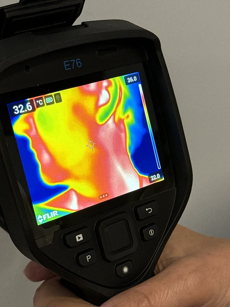
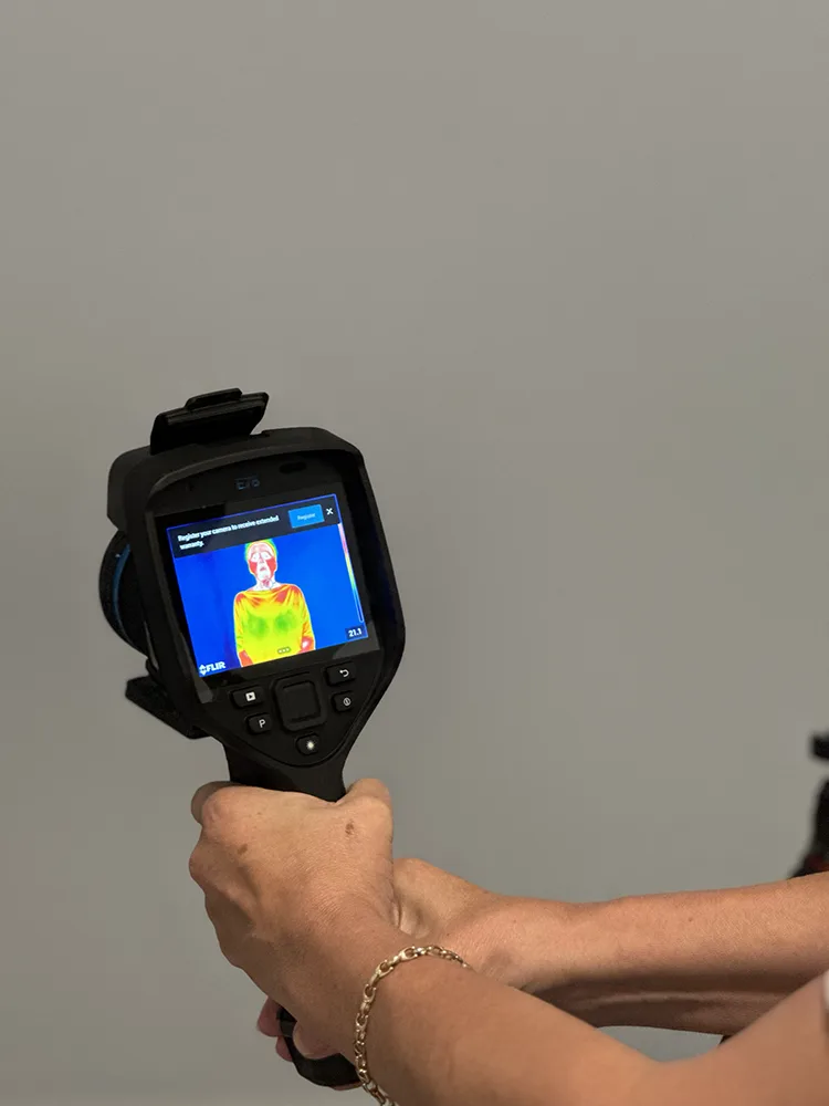
Medical thermography (DITI - Digital Infrared Thermal Imaging) is a non-invasive imaging examination using infrared radiation naturally emitted by the human body. Using a specialised thermovision camera, it is possible to analyse temperature distribution on the body surface and assess changes related to the physiological activity of tissues.
Every biologically active tissue produces heat as a result of metabolic processes and circulatory system function. Under normal conditions, the body maintains characteristic thermal symmetry. When disorders occur, such as inflammation, overload, microcirculation disturbances or increased metabolic activity, the temperature distribution may change.
Thermography records these changes in the form of a colour image called a thermogram. Individual colours correspond to specific temperature ranges:
- cooler areas are usually visible as shades of blue and green,
- warmer areas - as yellow, orange and red.
The examination is completely contactless and does not use ionising radiation.
Thermography is a functional examination
Thermography does not assess the anatomical structure of organs and tissues, as is the case with ultrasound, magnetic resonance imaging (MRI), computed tomography (CT) or X-ray.
The examination provides information about the physiological activity of the body, including:
- inflammatory processes,
- microcirculation,
- metabolic activity,
- thermal asymmetries,
- vascular tissue reactions.
For this reason, thermography complements imaging diagnostics and is always interpreted in the context of the patient's symptoms and other tests.
In what situations is medical thermography used?
Medical thermography is used as a complementary examination in assessing inflammatory processes, microcirculation disorders and changes in metabolic activity of tissues. The examination is used in neurology, orthopaedics, breast diagnostics, rehabilitation and monitoring of the body's regenerative processes.
Chronic pain and overloads
Chronic pain is not always associated with a visible structural change in imaging tests. Thermography can support assessment of tissue inflammatory activity, myofascial overloads and microcirculation disorders related to chronic tension or regenerative processes.
The examination is used for:
- spinal pain,
- muscle overloads,
- post-injury conditions,
- rehabilitation monitoring,
- chronic pain conditions.
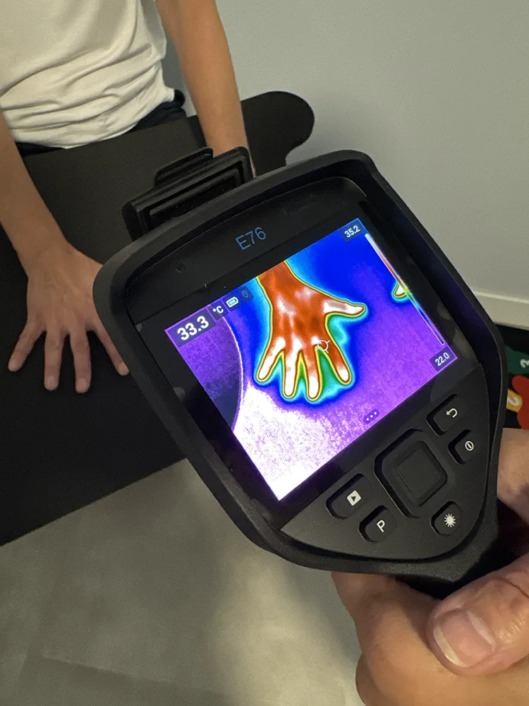
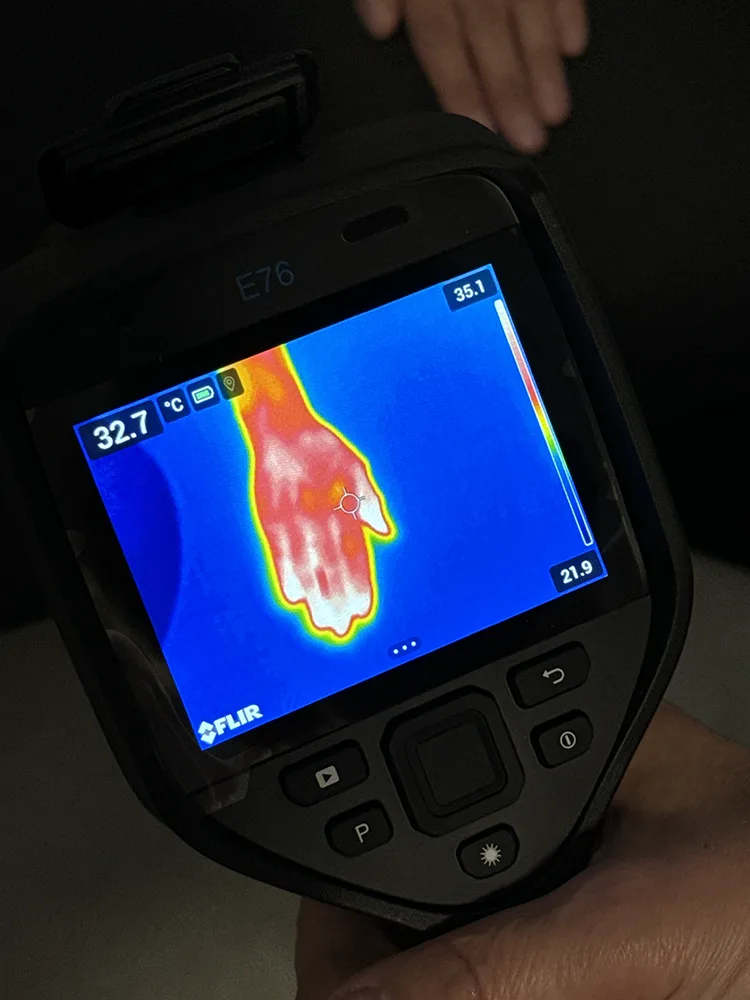
Circulation and microcirculation disorders
Changes in limb temperatures may reflect blood flow and microcirculation disorders. Thermography is used as an examination supporting the assessment of vascular problems and thermal asymmetries related to tissue blood supply.
The examination is used for:
- feeling of cold hands and feet,
- chronic swelling,
- Raynaud's syndrome,
- venous insufficiency,
- diabetic angiopathy.
Neurology and nerve complaints
Nerve conduction disorders and chronic pain syndromes can cause characteristic thermal changes visible on thermograms. Temperature asymmetry analysis is sometimes used as an element supporting functional assessment of the nervous system.
The examination is performed for:
- sciatica,
- neuropathies,
- neuralgias,
- carpal tunnel syndrome,
- chronic limb pain.
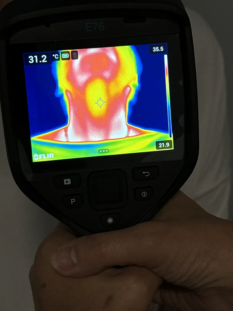
Thyroid and neck area
The thyroid is one of the most metabolically active organs in the body, so changes in its function can affect local temperature distribution. Thermography is used as a complementary examination in assessing the inflammatory and functional activity of the thyroid gland.
The examination can support diagnostics for:
- Hashimoto's disease,
- Graves' disease,
- thyroid inflammatory conditions,
- therapy monitoring,
- observation of functional changes.
Breast thermography
Breast thermography is a non-invasive functional examination supporting assessment of changes related to vascularisation and metabolic activity of breast tissue. The examination does not use ionising radiation and can complement imaging diagnostics.
Breast thermography is performed for:
- young women,
- women with implants,
- during menopause,
- as an element of change monitoring,
- as a complementary examination.
Rehabilitation and sports medicine
Thermal asymmetries may appear in areas of overloads, microinjuries and active regenerative processes. Thermography is used in rehabilitation monitoring and assessment of tissue readiness to return to physical activity.
The examination is used for:
- after sports injuries,
- with muscle overloads,
- during rehabilitation,
- in regeneration monitoring.
Chronic inflammatory conditions of the body
Thermal changes may accompany active inflammatory processes in various areas of the body. Thermography supports the assessment of physiological tissue activity and can be part of broader diagnostics of chronic ailments of ambiguous nature.
Frequently asked questions about medical thermography
Is medical thermography safe?
+
Yes. Thermography is a non-invasive and contactless examination. The thermovision camera does not emit radiation - it only records natural infrared radiation emitted by the body.
Does thermography use radiation?
+
No. Unlike X-ray or computed tomography, thermographic examination does not use ionising radiation.
What does the thermographic examination look like?
+
The examination is performed using a specialised thermovision camera from a distance of several dozen centimetres. Before taking images, the patient undergoes an acclimatisation stage in a controlled room temperature. Then a series of thermal images are taken and analysed by a specialist.
How long does the examination take?
+
The entire procedure usually takes about 30-45 minutes, depending on the scope of the examination.
Does the examination hurt?
+
No. Thermography is a completely painless and contactless examination.
How to prepare for thermography?
+
Before the examination, it is recommended to:
- avoid intense physical exercise,
- refrain from sauna and hot baths,
- not apply creams or ointments to the skin,
- remove jewellery from the examined area.
Detailed recommendations are provided before the visit.
Can thermography be performed during pregnancy?
+
Since the examination does not use ionising radiation, thermography can also be performed on pregnant women. Qualification for the examination is always decided by a specialist.
Who interprets the examination results?
+
The analysis of thermograms is performed by a specialist with appropriate competences in the interpretation of thermal images.
Current scientific publications on medical thermography
The development of infrared imaging technology and data analysis systems means that medical thermography is becoming an increasingly precise tool supporting the assessment of physiological processes in the body.
Contemporary research also analyses the potential of using artificial intelligence (AI) in the interpretation of thermographic images and supporting clinical diagnostics.
- Bauer, C. M., Rausch-Osthoff, A. K., Müller, B., et al. (2023). Use of infrared thermography in medical diagnosis, screening, and disease monitoring: A scoping review. Medicina, 59(12), 2139. https://doi.org/10.3390/medicina59122139
- Chen, D., He, L., Yang, J., et al. (2025). Infrared thermography in clinical practice: A literature review. European Journal of Medical Research, 30(1), 45. https://doi.org/10.1186/s40001-025-02278-z
- Lahiri, B. B., Bagavathiappan, S., Jayakumar, T., & Philip, J. (2012). Medical applications of infrared thermography: A review. Infrared Physics & Technology, 55(4), 221–235. https://doi.org/10.1016/j.infrared.2012.03.007
- Nallathambi, N., Bisaralli, R., Naidu, S. P., et al. (2025). Clinical application of infrared thermography in rheumatic diseases: A systematic review. Mediterranean Journal of Rheumatology, 36(1). https://doi.org/10.31138/mjr.271024.ita
- Branco, J. H. L., Branco, R. L. L., Siqueira, T. C., et al. (2022). Clinical applicability of infrared thermography in rheumatic diseases: A systematic review. Journal of Thermal Biology, 104, 103172. https://doi.org/10.1016/j.jtherbio.2021.103172
- Chantasartrassamee, P., Ongphiphadhanakul, B., Suvikapakornkul, R., et al. (2024). Artificial intelligence-enhanced infrared thermography as a diagnostic tool for thyroid malignancy detection. Annals of Medicine, 56(1), 2425826. https://doi.org/10.1080/07853890.2024.2425826
- Comparative evaluation of infrared thermography and mammography in the detection of breast cancer. (2025). Cureus. https://www.ncbi.nlm.nih.gov/pmc/articles/PMC12888057/
- Wojciechowska, A., & Serafin, Z. (2025). Medical thermography in the diagnosis of pressure ulcers: A narrative review. Journal of Wound Management, 26(2). https://doi.org/10.35279/jowm2025.26.02.07
- Mikulska, D. (2006). Współczesne zastosowania obrazowania podczerwonego w diagnostyce medycznej. Annales Academiae Medicae Stetinensis, 52(1), 35–40. PMID: 17131845.
Book thermography by phone or email

Next step
Start with a consultation.
Let's talk about symptoms, questions, and health goals. Together we will choose the right diagnostics or therapy.